| ■ Where is an eye (oculus) from? ■ |
| |
| ◆How were man’s eyes formed ?◆ |
It can be said that generating of eyes, i.e., the beginning of an organ of vision, started since a living thing began to have appeared on this earth.
Even a small living thing (monadic) without eyes can understand the light and the darkness. Moreover, although there is nothing that is called eyes in an earthworm, it has a cell (light sensation cell) which feels much light in the skin, and has caught a slight change of brightness well. Man's eyes could be called the result of evolution of such sensory cells. The present human being's eyes are that the fragment of cerebral born was divided from the remaining portion, and changed to the eyes gradually.
That is, probably, man's eyes may be called brain which jumped out. |
| Reference http://www.ocular.net/jiten/jiten001.htm
|
| ◆Structure of an eye◆ |
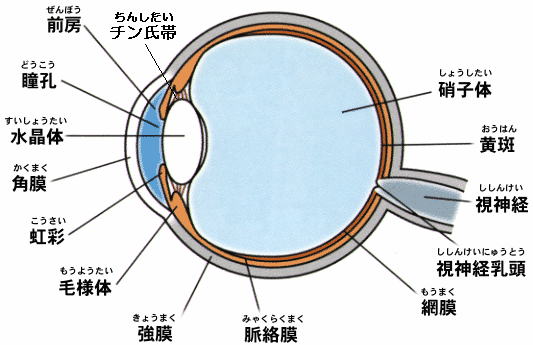
チン氏帯:Zinn's zonule、前房:anterior chamber、瞳孔:pupil、水晶体:crystalline lens、角膜:cornea、虹彩:iris、毛様体:corpus ciliare、強膜:sclera、脈絡膜:choroid coat、網膜:retina、視神経乳頭:optic disk、視神経:optic nerve 、黄斑:yellow spot、硝子体:vitreous body |
| 参照 http://homepage2.nifty.com/kaitsuka/index.html |
| ●Ciliary body |
It is the tissue with many muscles, and which carries out the focusing called regulation. |
| ●Iris |
The light volume which enters in an eyeball is adjusted in the brown portion in the surroundings of the iris of the eye. If it compares to a camera, it will be in charge of "an iris diaphragm." |
| ●Pupil |
It is a dark-eyed part. It is a way as light. It is in the front of a cornea eyeball and the curve has projected strongly from sclera. It is transparent and occupies one fifth of the outer membrane of an eyeball. |
| ●Crystalline lens |
The shape of a convex lens is carried out, and it is transparent and elastic. It coordinates with a ciliary body and performs the focus slide. |
| ●Vitreous body |
It is the portion surrounded by the crystalline lens and the retina, and
is choked up with transparent and the gel-like liquid. |
| ●Optic nerve |
the information from the visual cells of the retina is given to the lobe of cerebrum of a cerebrum -- so to speak, it is a signal cable. |
| ●Macular region |
The visual cells which manage sense of color and a sense of form are concentrating, and it is the sharpest portion of eyesight.
It becomes a portion which the light from the front forms the images. |
| ●Sclera |
The outer membrane of an eyeball is formed with the cornea. It is a portion called the white of the eye , and made of the very tough organization. |
| ●Choroid coat |
It is a membranae between sclera and the retina and the blood vessel manages many nutrition of the eyeball. Moreover, the duty of the blackout curtain is also carried out so that light may not enter from other than a pupil.
It is inside a retina choroid coat and is equivalent to the film as used in the field of a camera. Light is changed into electrical energy by visual cells, and it sends to an optic nerve. |
|
| ◆Comparison with a camera◆ |
| Camera |
→ |
eye |
| Body |
→ |
Sclera (white part of the eye) |
| Filter |
→ |
Cornea (black part of the eye) |
| Lens |
→ |
Crystalline lens |
| Iris diaphragm |
→ |
Luster |
| Film |
→ |
Retina |
|
|
With the latest camera, it can copy now easily, without putting a focus together.
This is because the image is tied with the suitable brightness for a film, while an iris diaphragm becomes large automatically by the strength of light, or becomes small and adjusts light. Thus, the structure of eyes and camera is well alike. |
| However, generally, although the camera of the automatic iris diaphragm is also made of these days, since the refraction mosquito of the light of a lens does not change, in order to make an image tie on a film, with a camera, you have to move a lens forward and backward (a focus is together put by changing a focal length). |
|
The crystalline lens which commits a lens changes the refracting power of light, and makes an image tie by the eye on the retina. |
|
Moreover, a film leaves the energy of light, while it has been physical. But, in the retina, the information received from visual cells is used as meaningful "screen", and it sends to a brain, and negatives "will be developed" for the first time in a brain, and the route of "seeing" will be taken. |
 |
 |
|
As shown in the upper figure, the picture projected on the retina is upside-down, but a brain is thinking that this upside-down image is projected upside down again, and is looking at the thing of the external world as it is. |
 |
| For further details of eyes, please refer to http://homepage2.nifty.com/kaitsuka/index.htm |