

12 cranial nerves
Twelve pairs of cranial nerves that mainly control the head and face enter and exit the brain stem.
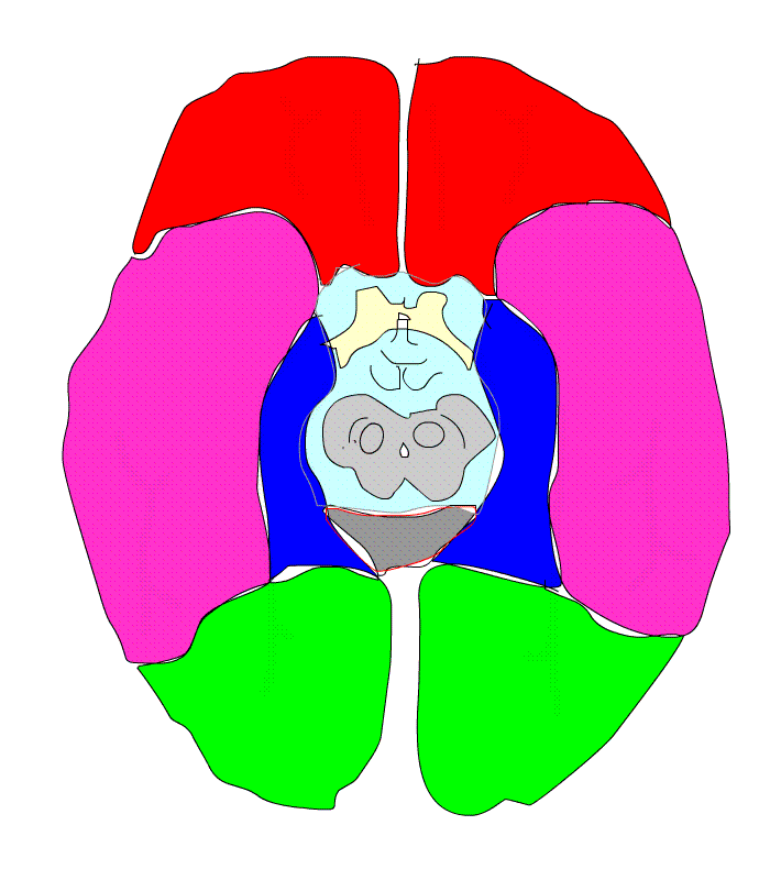
Let's look back, the brainstem is above the spinal cord, with the cerebrum above and the cerebellum on the back.
The brain stem is the area that combines the medulla oblongata, pons, and midbrain. Each area is embryologically different.
The medulla oblongata develops from ?mesencephalic vesicle, the pons from the ventral part of brain vesicle, and the midbrain from the mesencephalic vesicle.
Here let's learn about these cranial nerves.。
Quoted from https://nurseful.jp/nursefulshikkanbetsu/cranialnerve/section_1_00_01/
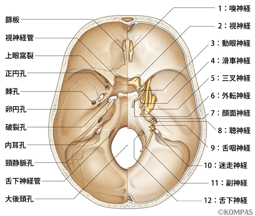
Quoted from http://kompas.hosp.keio.ac.jp/contents/000309.html
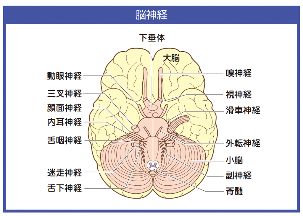
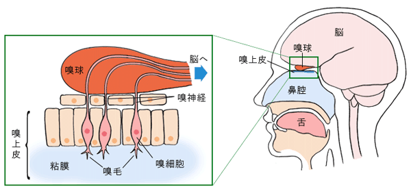
The figure on the left shows the brain from below. It is the view of the brain from the abdomen, which is the underside of the brain.
The animation below shows the figure 1 of structure of the brain and looks at the brain from ventral side.
The figure below shows the cranial nerve shown above exits the skull.
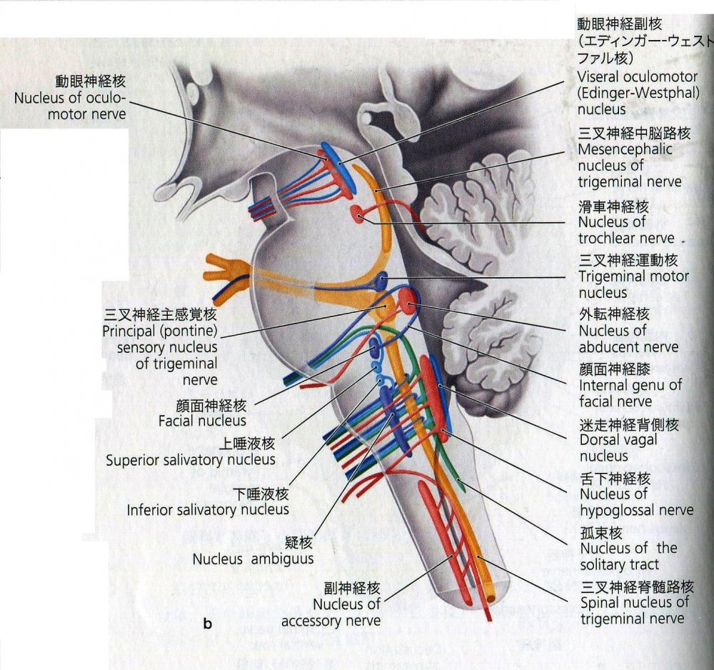
The figure below right shows the side of the cerebrum and is a side view of the midbrain, pons and medulla oblongata. It shows the location of the nucleus of the cranial nerve.
Watch where the cranial nerves go out from in the video.。
About the olfactory nerve
The olfaction is the most primitive sensory neuron.
The eye catches the stimulus from light .The ears catch the stimulus of sound.The skin catches the stimulus of the pressure and temperature. Unlike these, during the evolution of human beings, the olfactory sense was first developed to protect the body from the outside, and the taste sense was developed to check poison before eating.
How do you feel the smell? The sense is caused by molecules floating in the air or water. The place to receive this is at the top of the nasal cavity, which is called the chemoreceptor. When odor molecules stick to the receptors, electricity called the receptor potential is generated. During rest, only 5% of the air passing through the nasal cavity reaches the top. If you smell it consciously, 20% of the intake will reach the top.
In addition, the sense of smell and the sense of taste interact with each other during the diet. If you have a cold and clog up your nose, you may not be able to taste it or taste funny.
The amplitude, frequency, and duration of the receptor potential are transmitted to the center, and the type, size, and risk of stimulation are determined by the center.
Let's see the figure below.
Quoted from https://www.deodor.co.jp/nioimec.html
Olfactory cells are sensory cells that are replaced with new cells in about 30 days. There are many olfactory hairs at the edge of olfactory cilium.
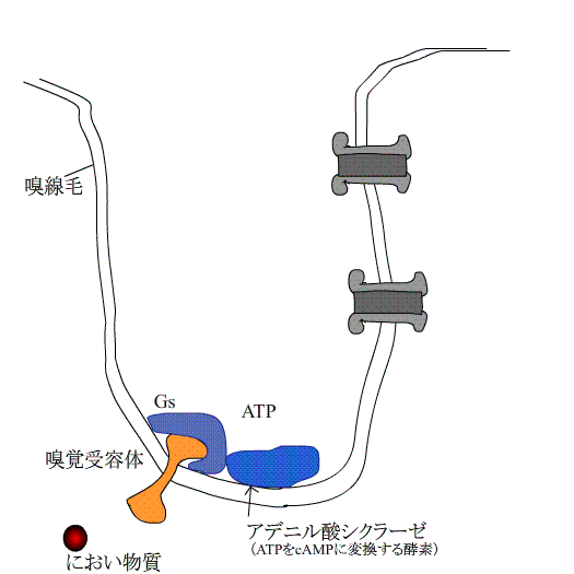
When an odorant sticks to the olfactory receptors on the olfactory cilia of the olfactory cell, Gs is stimulated, which activates adenylate cyclase.
cAMP is made from ATP. cAMP stimulates the channel, the channel opens, and Ca ions move inside the cell from outside the cell. At the same time, Na ions also move.
The increased Ca ions inside the cell stimulate different channels, and the CL ions inside the cell go out of the cell via the channel.
An electric potential is generated inside the cell due to the movement of such ions, and the stimulus is transmitted to the olfactory bulb via the olfactory nerve as an action potential.
One olfactory cell has several to dozens of receptor proteins. For this reason, there are olfactory cells that react to about 1000 different odorants.
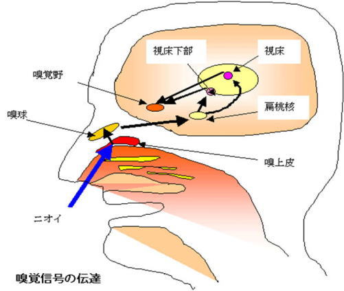
Communication with the center of the olfactory nerve
As shown in the figure below, the olfactory nerve passes through the olfactory tract and then connects to the ipsilateral and contralateral olfactory areas. Some reach the hypothalamus and trigger sexual behavior.
In addition, stimulation is transmitted to the hippocampus, amygdala, and limbic system. It also connects to the orbitofrontal cortex via the thalamus.
Determination of odor
Each olfactory cell responds strongly to particular odorants.
Thousands of odors have distinctive patterns that excite each olfactory cell, and the brain remembers and identifies odors with different patterns in the orbitofrontal cortex.
The garlic odor seems to be noticeable at a little concentration. The irritating odor of wasabi is used as a method to inform the hearing impaired of abnormalities such as fire. Since olfactory cells adapt quickly to the same odor, they are sensitive to the odor of others, but are insensitive to the odor themselves.
Humans are 1 to 10 million times less sensitive than dogs.
About the optic nerve |
First of all, Let's see the development of the eyes in the animation below.
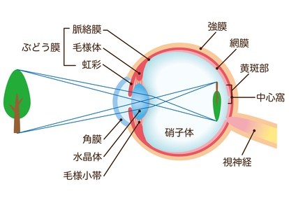
The eye is made from a neural tube called the forebrain. The eye is a part of the brain that seems to have popped out.
The cornea and lens originate from the same ectoderm.
The retina and pigment epithelium cells are made of the same nerve layer.

Let's review. External light or images pass through the cornea, lens, and vitreous body and into the inside of the retina.
What kind of cells process information in the retina?
Please see the animation below. The top is the inside of the retina, where the light is first received.
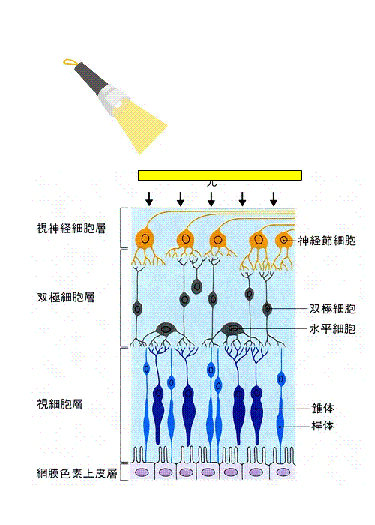
This side is the end of the retina (backside)
Light reaches the end (backside) of the retina first. There are two types of cells. As in the animation, they are pyramidal cells and rod cells.
Pyramidal cells with cone-shaped cells respond to strong light, increase the accuracy of resolution, and are involved in color vision. There are about 6 million of them in the center of the retina.
Rod cells with a columnar? tip have high sensitivity and respond to weak light . There are more than 100 million and they are located in the peripheral area of ??the retina.
Nocturnal cats have many rod cells, and most birds have pyramidal cells.
Bipolar cells receive signals from pyramidal and rod photoreceptor cells and transmit them to ganglion cells.
In the central part of the retina, pyramidal cells: bipolar cells: ganglion cells correspond to 1:1:1, indicating high resolution.
At the periphery of the retina, more than 100 rod cells correspond to one ganglion cell.
Ganglion cells are multipolar neurons. There are about 1 million large multipolar neurons. The cell body has several layers in the macula and one layer in other parts.
Multipolar neurons are shown in the figure below,
The number of protrusion is one...?unipolar neuron
The number of protrusion is two...?bipolar neuron
The number of protrusion is more than three...multipolar neuron
One multipolar neuron protrusion extends from the cell body, which is further Branching... pseudounipolar neuron
Here, cells have rough endoplasmic reticulum but axons and dendrites don't have it. This rough endoplasmic reticulum is a factory that makes proteins.
This is the differences between axons, dendrites and cell bodies.
Quoted from http://www.geocities.jp/ululu_o_ululu/report-07-01.htm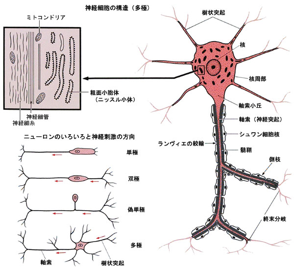
Next,
The horizontal cells seem to integrate the information from the photoreceptor cells by extending the protrusions that connect adjacent photoreceptor cells. The cells that fill the space between these neurons are called Muller cells. These cells make the retina a kind of closed environment, protecting the retinal neurons from harmful substances.
Retinal pigment epithelial cells maintain the function of photoreceptor cells.
1) Prevents the choroidal tissue fluid from entering the retina. Even if inflammation occurs in the sclera and choroid, it protects the retina.
2) It absorbs the light that did not react with photoreceptor cells and prevents scattering.
3) The material of the visual substance that absorbs light is supplied to the visual cells and the old substance is processed.
Retinal detachment is a condition in which the retinal nerve layer is detached from the pigment epithelium layer , and various substances cannot be received from the pigment epithelium cells, resulting in a loss of visual field
Quoted from http://www.takatsu-chiro.com/yougoshu/cranial-n-2.htm
"Medicine for children" page http://www.skmc.jp/anime/child/eye/eye_index.htm
The chapter "What is the eye?" in the website above, what you are looking at is upside down on the retina.
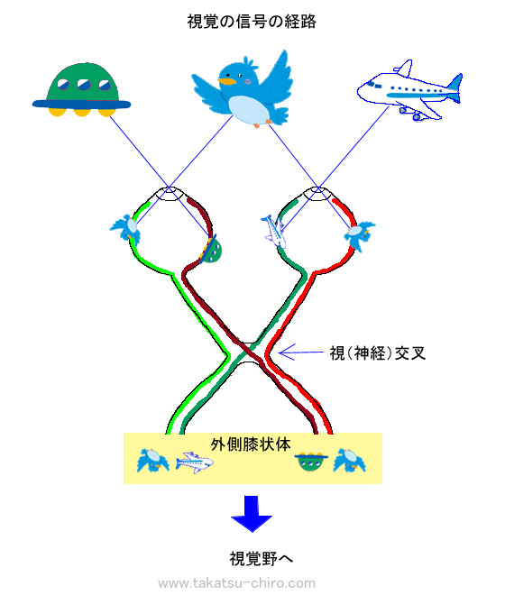
Most of the optic nerve exiting the eye passes through the optic chiasm and becomes the optic tract, ending in the lateral geniculate body of the thalamus.
The lateral geniculate body that holds the optic nerve together is called the relay nucleus of the optic nerve.
The lateral geniculate body has two layers, a large cell layer and a small cell layer, and the large cell layer is sensitive to moving light spots. The small cell layer responds to persistent light. In other words, information about movement and information about detailed shapes and colors are relayed separately, become visual rays, and are communicated to the primary visual cortex.

In the animation on the left,you acn see the information from the visual line goes to the primary visual cortex.
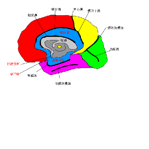
Where is this primary visual cortex?
Please see the picture below. It is in the?calcarine sulcus.
| How is visual information processed? |
In the cerebral cortex, the area that processes visual information is about 1/3 of the total area .
Sight 87% Hearing 7% Tactile 3% Olfactory 2% Taste 1%. Actually, we don't judge everything by our eyes, so in some cases, we think that information other than eyesight also affects the cerebrum a lot. However, there is no doubt that the amount of visual information is huge.
So how is visual information processed?
In the primary visual cortex,
Cells that respond to elongated slit-shaped light directed in a specific direction. - Simple cells.
Cells responsive when slit light moves in a specific direction-Complex type cells.
Cells with slit shaped light with specific length and direction-Dobble complex cells.
It seems that these three cells decompose visual information into elements such as the inclination, length, and moving direction of line segments to create the outline of an object
It has been explained that simple and complex cells that are restricted to the same direction gather and react in the vertical direction, but it is quite difficult.
What we know is shown below.
The primary visual cortex is above and below the?calcarine sulcus . Visual information from the primary visual cortex is analyzed in the previsual cortex, which is located in front of the primary visual cortex . After that,the visual information is sent to the visual association cortex of the temporal lobe and parietal lobe . It seems that in the temporal association area, information on shape and color is integrated to perform object recognition of what the object is. In the parietal association area, spatial recognition is performed based on movement and depth information . The temporal lobe and parietal lobe on the right side are related to the visual information that enters from the left eye, and the temporal lobe and parietal lobe on the left side are related to visual information from the right eye, so obstacles such as shape, color, and movement are shown at opposite side. (The obstacles is explained at chapter No. 8 movement mechanism) |
The story so far is that the light reflected on the retina is converted into an electric signal, and the action potential, which is an electrical spike, is transmitted from the retina through the optic nerve, to the occipital lobe, temporal lobe, and parietal lobe and the image was recognized. The scenery that is analog becomes digital in the retina and is recognized again as analog in the cerebrum.
In the outer segment of photoreceptors, light turns into electrical signals.
Let's talk about photoreceptor cells at the next Chapter.