

The blood vessel at brain
The point of this page The main blood vessel for the cerebrum is the internal carotid artery. The blood vessels for the brainstem and the cerebellum are vertebral artery and basilar artery. The brain consumes the biggest energy in the organs. There are 2 kinds of cerebral hemorrhage, a subarachnoid hemorrhage and a brain hemorrhage. |
You can see how the blood goes from left ventricle to the brain.
The ascending aorta comes from the left ventricle and become brachiocephalic artery on the right side. The brachiocephalic artery is separated to right common carotid artery and right vertebral artery.
The right common carotid artery is separated to external carotid artery and internal carotid artery at the neck. The left common carotid artery is separated same.
The arterial blood goes to the brain through the internal carotid artery.
The both sides of the external carotid artery mainly goes to the face and the dura mater. The vertebral artery goes besides the spinal cord thorough foramen magnum to the brain.

Cross-section from the front face
The brachiocephalic artery exists only on the right side. The right brachiocephalic artery is connected to the right common carotid artery.
The vertebral artery from the subclavian artery goes to the brain from the backside of the head.
You can see the arterial blood is flowing internal carotid artery, deutocerebrum, protocerebrum, postcommunical part in this left animation.
Both side of the vertebral artery connect at the basilar artery and flows to the cerebral artery.
the basilar artery is separated to arteries related to the brainstem and cerebellum.
copyed from http://www.yamamura-clinic.net/index.php?id=73
where is the place the main artery running?
First of all,here I explain a cross section of Cerebrum.
To quote from https://ja.wikipedia.org/wiki/%E5%86%A0%E7%8A%B6%E9%9D%A2

The sagittal plane is the section from the side.
coronal plane is the section from the front.We call it frontal plane as well.We use it for spilt the body to the ventral side and the dorsal side.
The below figure shows the transverse plane.
To quote from https://rehatora.net/%

The below figure shows the sagittal plane.
to quote from https://rehatora.net/%
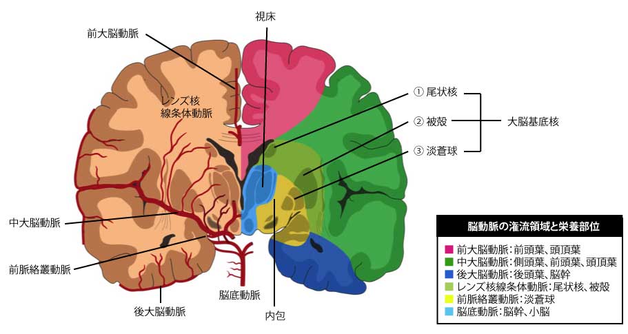
Each of blood vessel region has each work.When a clot of blood blocks the vessel,many obstacles happen.
Let's talk about the cerebral hemorrhage. There are 2 kinds of it,subarachnoid hemorrhage,and brain hemorrhage.
Subarachnoid Hemorrhage
the figure below shows, the blue is the dura mater,the yellow is the arachnoid,the white is subarachrnoid cavity.
In the subarachrnoid cavity,There is the artery floating in the cerebrospinal fluid.
The hump called aneurysm sometimes happens in an artery, though the reeason is not clear.
When the hump is broken, the blood goes into the subarachnoid.This is how subarachnoid hemorrhage happens.
cerebrospinal fluid is dyed with bloods.Check the animation.

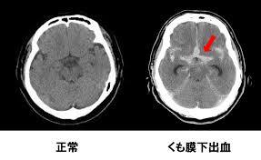 The watery stuff is hown white in CT.The cerebrospinal fluid is located at the subarachnoid around the brain and is shown like the top right figure.
Brain Hemorrhage
As you see in the below animation,the thick vessel goes to thin vessel at the brain tissue.
When the vessel is broken,it bleeds.
cerebrospinal fluid at the subarachnoid is hardly mixed with blood as shown in the below animation.

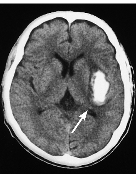
The blood is hardly seen in the brain,so you can see the white blood lump clearly as shown at the right figure.However when the amount is too little and the white color is too pale,it is failed to be seen.
http://www9.plala.or.jp/sophie_f/disease/cerebral4.htmlから引用
The vessel causing subarachnoid hemorrhage is the vessel floating in the cerebrospinal fluid. The vessel causing brain hemorrhage is the thin vessel at the brain tissue. This is the reason the bleeding in the brain is seen in the different way. |
次に、
| How much blood flows to the arteries in the brain? ? ? |
The brain has?a?mechanism that keeps?blood flow constant regardless of changes in blood pressure?.
As in the animation on the left, when the average blood pressure (= minimum blood pressure + (maximum blood pressure-minimum blood pressure) / 3) is in the range of 60 mmHg to 150 mmHg, the cerebral blood flow is 50 to 55 ml / min / 100 g of brain dry weight.
A mean blood pressure of 60 mmHg probably corresponds to a systolic blood pressure of 75 to 70 mmHg and a diastolic blood pressure of 55 to 50 mmHg.
A mean blood pressure of 150 mmHg probably corresponds to a systolic blood pressure of 210 to 200 mmHg and a diastolic blood pressure of 130 to 120 mmHg.
| 血The blood flow in the brain does not change even when the?blood?pressure is low or significantly high, so you can think without fluttering or headache. |
But, surprisingly, the cerebrovascular system is affected by the amount of oxygen and carbon dioxide dissolved in the arterial blood. The above-mentioned brain blood volume changes extremely.
Human beings, animals, eat and convert food into nutrients in the body with various substances called enzymes.。
At this time, oxygen is used, and carbon dioxide and water are formed in the rest. Carbon dioxide is dissolved in the blood. This carbonic acid is referred to as PaCO2.
The normal value of PaCO2 is in the range of 43 to 47 mmHg.?If you are not sick, you will be adjusted within this narrow range.It's amazing
では、下に炭酸ガスにThen, the figure of the state of the cerebrovascular where it responds to carbon dioxide is shown below.?The red line is the cerebrovascular response to blood pressure, as mentioned above.
The blue line is the reaction to carbon dioxide.?The horizontal axis represents the change in carbon dioxide.?The unit is mmHg.
Regardless of blood pressure fluctuations, when carbon dioxide in blood becomes higher than the normal value, cerebral blood vessels open, a lot of blood flows in the blood vessels, and blood flow increases.
When it is lower than the normal value, cerebral blood vessels contract and blood volume decreases.。Carbon dioxide has a stronger effect on cerebral blood vessels than fluctuations in blood pressure.
Next, let's consider the effect of oxygen in the blood on cerebral blood vessels.
Human arterial blood contains a large amount of oxygen obtained from the lungs, and after oxygen is used in each tissue, venous blood returning from the tissue is low in oxygen. Oxygen dissolved in arterial blood is expressed as PaO2. a is the a in artery.
In the case of venous blood, it is expressed as PvO2.?P is pressure and v is vein's v in English.?O2 is oxygen.
The normal value of PaO2 in arterial blood is 95-98 mmHg.
When PaO2 falls below 50 mmHg, the cerebral blood vessels expand at once and the blood flow in the cerebral blood vessels increases.
Unlike carbon dioxide, blood volume does not change even if oxygen increases above the normal level.?that's strange.
For humans, when oxygen in arterial blood is low, they cannot live.?In particular, the brain is vulnerable to oxygen.
If PaO2 is below 50 mmHg, the brain will be tough.?Therefore, even if blood is low in oxygen, a lot of blood will be carried to the brain.?Therefore, the blood vessels of the brain open.Blood vessels open faster than blood pressure changes, and blood vessels open in response to changes in oxygen.
next,
Considering the blood flow of the whole body, in the case of an adult weighing 65 kg,
How much blood (ml) circulates in one minute
Looking at each organ,
organ rest exercise Comparison
| Lungs? | 5000 | 25000 | About 5 times |
| brain | 650〜750 | 750〜1000 | About 1.1 to 2 times |
| Gastrointestinal / liver | 1000〜1300 | 750〜1300 | About 3/4 ~ equivalent |
| kidney | 1000 | 500〜1000 | About 1/2 to equivalent |
| muscle | 750〜1000 | Approximately 15 to 22 times total for muscle and skin | |
| Skin | 150〜300 | 20000〜21000 | |
| Bone / fat | 500〜750 | 250〜500 | About 1/2 to 2/3 times |
Blood flow in the lungs is the amount of blood that comes directly from the heart.00
After that,the blood splits and flows to various organs.
The amount of blood in the lungs is the same as the sum of blood distributed to each organ.
During exercise, the blood flow in the skin and muscles increases about 20 times, and unexpectedly the brain also slightly increases.
The amount of blood flow in the brain is 15% of the blood flow from the heart
Brain energy consumption per organ weight is the biggest in the body
Brain weight is only 2% of body weight, but its energy consumption is 18% of the body.
| Organ | Energy consumption (%) | Organ weight (%) | Energy consumption per weight (Kcal) |
| brain | 18 | 2 | 900 |
| heart | 11 | 6 | |
| kidney | 7 | 633 | |
| liver | 20 | 52 | 48 |
| muscle | 20 | ||
| Skin | 5 | ||
| Other | 9 | 40 | 48 |
The total organ weight of the heart and kidneys is 6%,Energy consumption per weight is 633 Kcal
52% for liver, muscle and skin.?48 Kcal.
Even when you are still, only your brain uses a lot of energy.
So what is the source of brain energy???????
Mainly glucose, that is glucose.? When glucose is not available, such as when the disease is severe, a substance called the ketone,body of the fat componenent,is also used.
The brain always consumes 46% off the total body glucose consumption.?When we sleep, we use 5g of glucose per hour.?(A person consumes glucose about 260g in total without doing anything a day. 120g of this is used by the brain .) |

This?is called the?Blood Brain Barrier?.
 In the case of a normal blood vessel, various substances leak from the blood vessel to the cell via the Pore.
Unlike the capillaries other than the brain, the capillaries in the brain do not have a Pore.So,substances cannot freely enter or leave to the brain.?By preventing the substances in the blood from easily passing to the brain,the capillaries plays a role in protecting the brain from toxic substances.?On the other hand,medicine cannot easily pass through either.
Lipid soluble (ketone body of lipolysis product) and glucose aa (amino acid), which are energy sources of nerve activity, can pass through.。Alcohol and narcotics contained in alcohol and drug are all fat-soluble, so they can penetrate into the brain, which is rich in fat.
The figure on the left is taken from http://www.js-brain.com/glial.html
About the venous system of the brain
The picture on the left shows the main venous system.
It is divided into four.
Cortical vein system that runs on the surface of the brain
Medullary vein system running in the parenchyma
Subependymal venous system running under the ependymal ventricles
Meningeal veinin the dura working for final venous return
Quoted from http://www.geocities.jp/study_nasubi/j/j12.html
Please watch the anime on the left slowly.?The color of blood vessels changes.
Restructured from http://chousei58.com/?p=40052
The blood vessels distributed on the face and the side of skull,flow to the?external jugular vein which is shown as dark blue in figure.
Venous blood from the brain in the skull
gather in various?sinuses
It flows into the internal jugular vein,
which is shown as bright blue in figure.
Cerebral arteries do not flow besides veins in other organs.
the presence of a sinus is unique to the brain,it doesn't be seen in other organs.
What is a sinus???
Before that, the structure of veins and arteries is shown below.
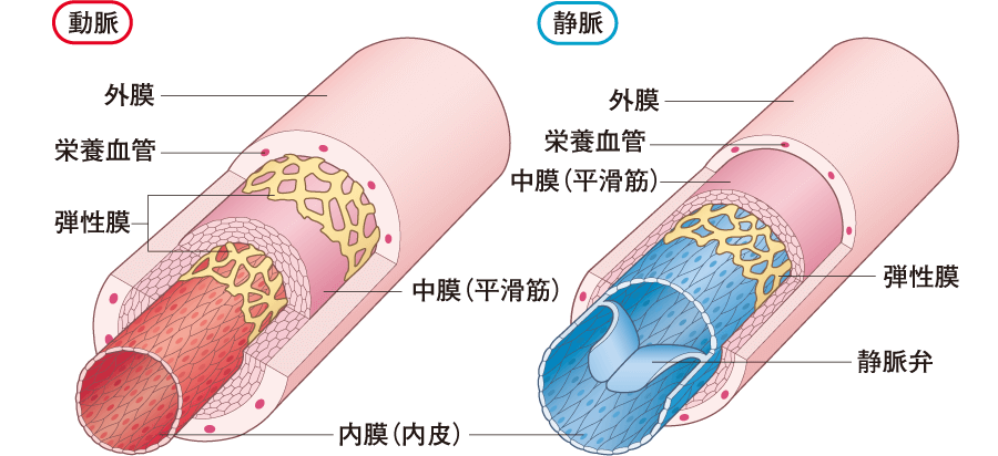 The artery has a?thick?elastic membrane and?has a stretching force.?Veins are less elastic than arteries.?The vein below the heart has a venous valve.?This is because it returns blood to the heart.?The veins above the heart have no valves.?That is, there are no valves in the cerebral veins.?The figure on the left is taken from https://www.kango-roo.com/sn/k/view/1840
では静脈洞とはどういう構造でしょうか。
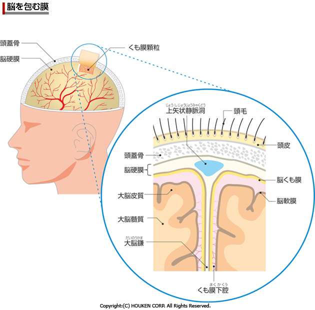
As shown in the picture on the left, the blue part represents the sinus.?The superior sagittal sinus is one example.
The outer and inner lobes of the dura are usually in close contact with each other, but in some places such as the midline, there is a gap between the outer and inner lobes, where venous blood gather. This spot is called?the dural?sinus?.?Reprinted from https://health.goo.ne.jp/medical/body/jin009
Venous blood collected in the sinus flows into the left and right internal jugular veins,
Venous blood from the face, etc. flows into the left and right external jugular veins, which become the left and right brachiocephalic veins, flow into the superior vena cava, and go to the right atrium
Return to the first page. Go to the web for kids learning medicine.