

Let's talk again about the cerebral neocortex.
At first glance, the cerebral cortex looks same in every part, but each part has completely different functions. This is called functional localization.
In 1861, Broca,a scholar examined a patient with motor aphasia and found that there was damage to the posterior inferior frontal gyrus ( Broca's area ) in the left hemisphere.
The patient with motor aphasia can listen to and understand what others say, but can't speak or hardly speak.
Wernicke,a scholar found sensory aphasia in a patient with a disrupted posterior superior temporal gyrus.This area is called the Wernicke field .
The patient with this damage can be talkative and speak fluently, but the contents are almost meaningless. they can't understand what others say.
Next,
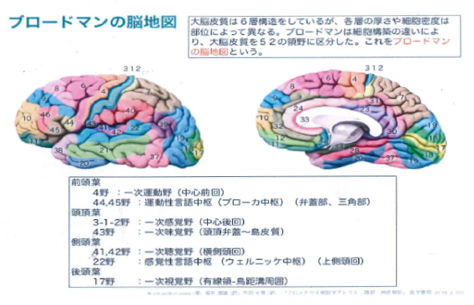
German neuroscientist Korbinian Brodmann was focused on the method to stain the different types of cells of the brain that has been developed by Franz Nissl. Since each layer has different thickness and cell density, he classified those and in 1909 the human brain was divided into 52 different areas.This division is called the Broadman Brain Map.It was very accurate , such as the division of areas 17 and 18 in the visual cortex.
Well then, What happens when the cerebral cortex is stimulated with a weak electric current?
It induces limb movements and causes muscle contraction, as in the animation below.
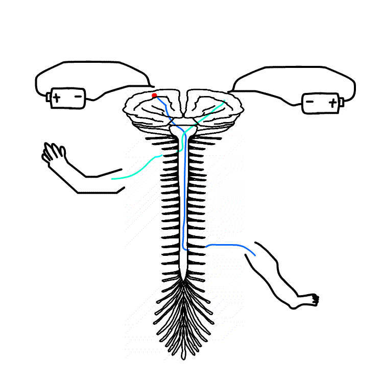
The places where you can induce movement are shown in animation of the side view of cerebrum below.
It is limited to the precentral gyrus just in front of the central sulcus. It is called the primary motor cortex.
Next, as in the animation below, when the postcentral gyrus located just behind the central sulcus is stimulated with weak electricity, feeling of contact and pressure on hands and feet happens. It is not a strong pain.
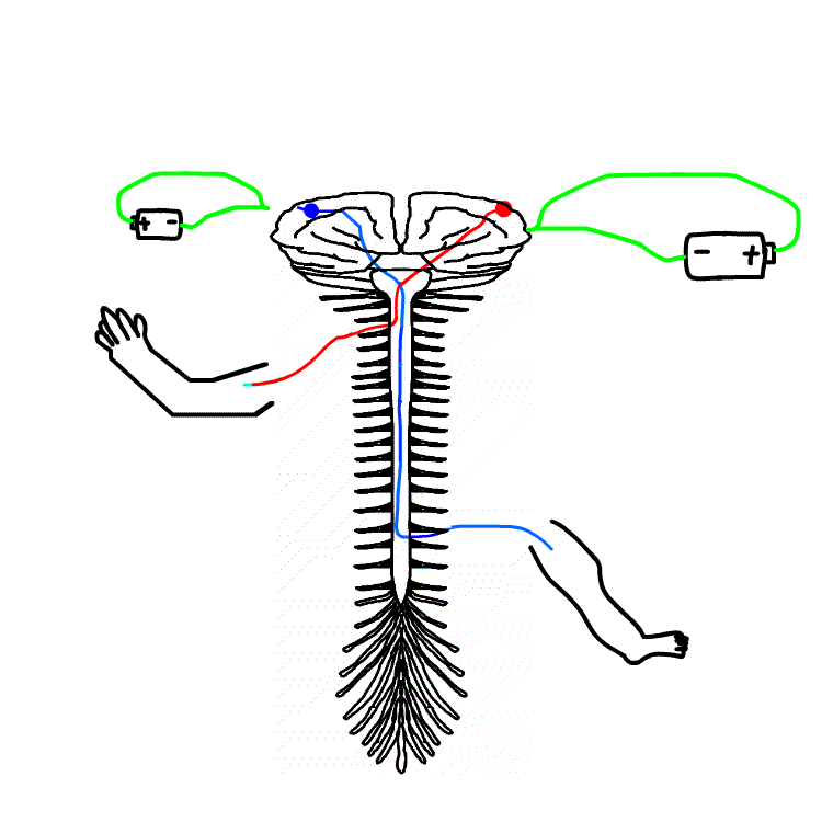
When we are pinched, it hurts and we feel pressed. At this time,the stimulus from the skin passes through the nerve,goes to the spinal cord and the cortex of the cerebrum,and causes pain or pressure.
In this experiment,we applied a weak electrical stimulus to the cerebral cortex from the opposite direction that the stimulus is transmitted through nerves. By this way,we searhed where to react on the skin and made a map of perception in the cortex.
What is somatic sensation?
There are?sensations?that can be caused by other than?the?sensory organs?such as the eyes, ears, nose, and tongue?.
In other words,the?sense on the skin?, such as sense of touch?,?pain?or the deep sensory?such as muscle?contraction?, algesia caused by change of organ,such as headache, stomachache,and chestache.
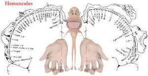
The location of this primary somatosensory is postcentral gyrus just behind the?central?sulcus?.
Penfield (1891-1976),Canadian brain surgeon,created a brain map showing which part of body correspond to the cerebrum.?The size of each body part and the size corresponding to the brain map are not in a proportional relationship.?In other words,hands and face move more finely than the feet, so the proportion of hands and face in the cortex is much wider than that of the feet.?The same can be said for the senses.

A model in which the size of each part of the body is replaced by the size on the brain map is called "homunculus".
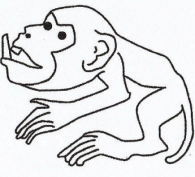
| Figure3 Animal Homunculus | ||
| monkey | cat | rabbit |
 |
 |
 |
| Figure-3. From Yuji Iketani "Over-Evolved Brain" | ||
The part for whiskers of cats and rabbits is large, which means it is an important sensory organ. When I was a kid, I was told that I shouldn't cut the whiskers of a cat.


"Human Homunculus" has large tongue, lips, fingers (especially forefinger), and large eyes compared to the body.
The Homunculus, in the sense of dwarf in Latin,means Android and its technology made by Europian alchemist. It is used to explain the brain function by fitting parts of the body to the brain and comparing it to dwarfs.
As mentioned above, Broadman's brain map and Penfield's brain map are used together to study brain function.
Sensory area review
In addition to the primary somatosensory area, secondary somatosensory area and primary motor area, there are primary visual cortex and primary auditory cortex .
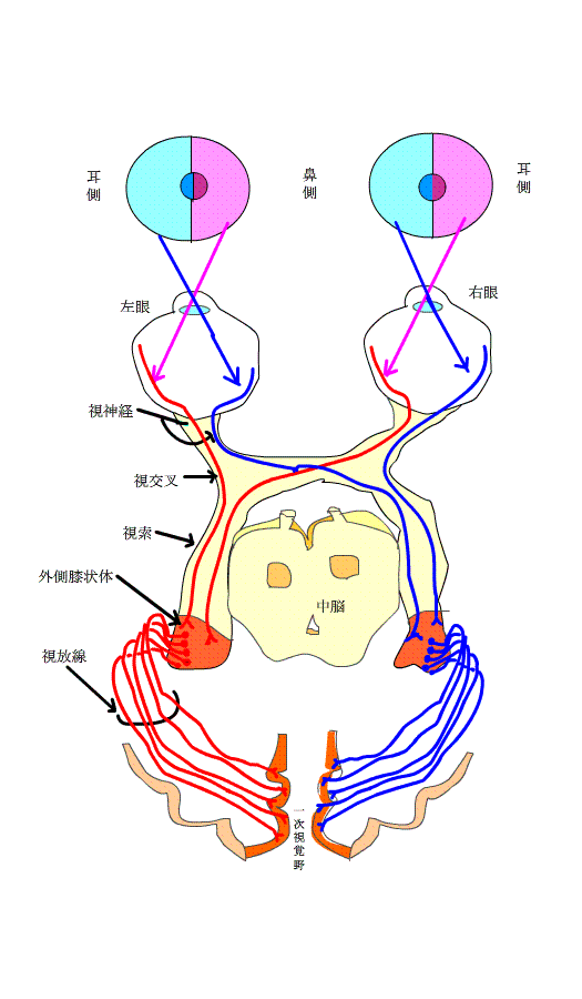
The primary visual cortex is on the medial side of the occipital lobe, above and below the calcarine sulcus (Brodmann17). The animation below shows that abnormalities in the visual field allow us to infer disorders of the optic nerve, optic chiasm, optic tract, and optic radiation.
next, I will explain about light reflection . When the retina catches bright light, the pupil contracts. This is called reflex.
The diameter of the pupil changes from about 8 mm to 1 mm in about 0.2 seconds, changing the amount of light reaching the retina up to 64 times.
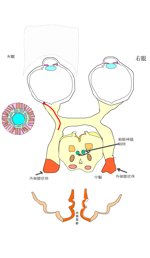
In the animation below, a slightly dark state is created in the left eye, and a flashlight is suddenly applied there. At this time, the stimulus sensed by the retina is divided into the route to the lateral geniculate nucleus and the route to the pretectum of the midbrain, as shown by the red line.
The path to the lateral geniculate nucleus goes to the primary visual cortex that identifies things, as in the animation above.
The stimulus goes both left and right preoptic region. From here, the stimulus is transmitted to accessory nucleus of oculomotor nerve.
As shown by the blue line, this stimulus goes ot the ciliary ganglion and travels to the musculus sphincter pupillae.
When the light from a flashlight is intensely applied to the retina of the left eye, both the left and right musculus sphincter pupillae contracts.
In other words, when the right eye is blindfolded and the left eye is exposed to light, the right eye also contracts.Even if the right eye does not receive light.?
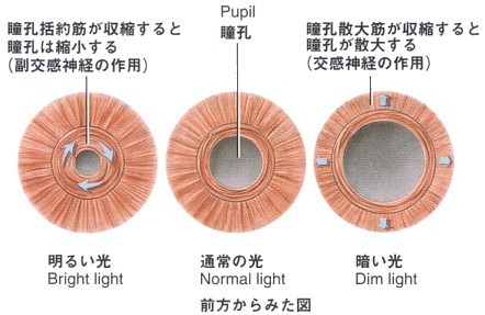
Then, the size of the pupil becomes smaller.This is called miosis.
Figure blow shows a brief dissection of the musculus sphincter pupillae.
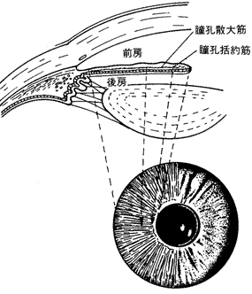

The musculus sphincter pupillae has a donut shape. When this muscle contracts, the donut shape becomes smaller.
When relaxed, the donut shape grows larger.
In the center figure below, it is the size of the pupil in normal light, but it is dazzling in bright places because a lot of light enters the retina. To reduce the amount of light, the size of the pupil becomes smaller, as shown in the bottom left figure.
At this time, the dilator pupillae muscle does not change much.
Then, what does the dilator pupillae muscle do?
When the amount of light decreases, that is, when it is dark, people try to open their pupils and capture a large amount of light into the retina so that they can see clearly.
At this time, not only do the musculus sphincter pupillae relax and enlarge the pupil, but dilator pupillae muscle contracts and the pupil becomes large in order to further enlarge the pupil. It shows in the right figure below.
There are other reflexes that cause the pupil to constrict.
They are accommodative reflex and convergence reflex.
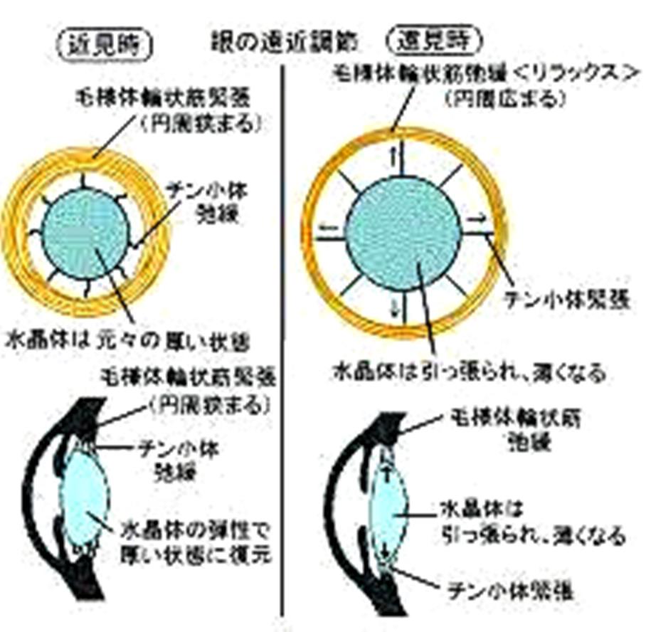
accommodative reflex
The lens swells when you look closer. The refractive power of the lens increases, making things easier to see.
See http://www.skmc.jp/anime/child/eye/eye_2.htm
At this time, as shown in the figure below, the ciliary circular muscle becomes tense and the circumference becomes tight.
The ciliary zonule become loose. Then, the lens that was pulled by the ciliary zonule returns to its original thickness.
Also, when you see far, the ciliary circular muscle becomes loose and the circumference widens. The ciliary zonule, on the contrary, becomes tense and pulls the lens outward. The lens becomes thinner, making it easier to see objects.
The miosis also happens at that time.
If you open your eyes (mydriasis), you will use the lens widely. Using a wide lens means that light rays come from many directions, so that the lens are focused only at a specific distance.
On the other hand, with miosis, only the central part of the lens is used. The light rays will only be received in parallel from the front, making it easier to focus. This means the depth of focus is big.
When people with nearsightedness try to see with the naked eye, the y sometimes narrow their eyes. This makes it easier to focus by further narrowing the eye diaphragm.
Like a pinhole camera, if you set it to an extreme aperture, it is called "pan focus" and it will focus at any distance.
Convergence reflex
When you gaze at an object at close range, the visual axes of both eyes get closer. This is called congestion. When you gaze at an approaching object, the muscles called the internal rectus muscles inside your eyes contract at the same time, forming a cross-eyed.
Postscript
There are five functional localizations in the cerebral cortex: motor cortex, somatosensory cortex (primary and secondary), language cortex (sensory and motor), visual cortex and auditory cortex.I haven't mentioned the auditory cortex this time.