

The motor center, which was important for sub-avian animals, was driven deep into the cerebrum, the medulla, in humans, whose cerebral cortex was highly developed.This is called the basal ganglia . This movement is performed by a neural circuit called the extrapyramidal system.
When humans voluntarily move, commands from the cerebral cortex (willingness to move) are transmitted to the spinal cord via nerves and move the muscles of the hands and feet. This neural circuit is called the pyramidal system, which is a special motor system that first appeared in mammals .
The basal ganglia, which is an extrapyramidal system used in sub-birds animals, completes movement from the pyramidal system smoother and more balanced in humans. There is no pyramidal tract in sub-birds animals, and the basal ganglia occupies the most important position in the motor conduction tract.
Whereas the pyramidal tract is an anatomical entity, the?"external pyramidal tract" neural tract does not exist anatomically?.?For this reason, in addition to the expression "?extrapyramidal symptom" in clinical medicine, the term extrapyramidal is being used less frequently as inappropriate
| The basal ganglia is an important area for the smooth movement of limbs. |
In humans, the movement disorders caused by the damage to the basal ganglia are roughly divided into two.
The first type is hypokinetic and hypertonia.The representative disease is Parkinson's disease.The other type is phyerkinetic / hypotonia.The representative disease is Huntington's chorea.
Parkinson's disease generally involves small movements of the limbs and stiff muscles, regardless of your will.The movement is slowed down and the facial expression disappears.
This is a video provided by Dr. Yasushi Miyagi, Functional Neurosurgery, Medical center Souseikai Fukuoka Mirai Hospital.
You can see the difference of tremer before and after the treatment method called subthalamic nucleus stimulation.
This is a video of a patient with severe body tremor.
The symptom of Huntington's chorea seems restless because of the quick movements of frowning and shrugging, regardless of your will.?It becomes stronger when walking, trying to do something, or getting tense than when resting.
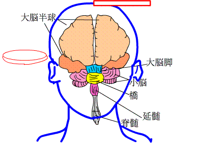
Now, as the cerebrum grows larger, the basal ganglia becomes smaller, and it is put inside the brain.Let's see it in anime.
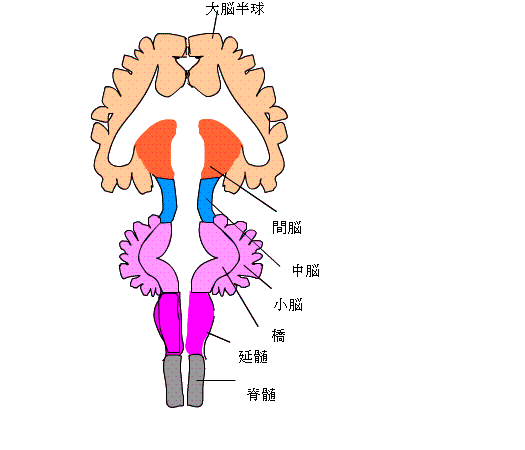
The cerebral hemisphere is larger and the diencephalon, midbrain and its surroundings are smaller
Let's see the horizontal section and the frontal section of the brain in animation.
 A horizontal cross section?crosses the brain shown as the red disk left in anime.
Frontal section vertically cuts brain from the top to bottom shown as the red rectangle on the right.
Next, I tried to show what you can see in these cross sections with an animation.
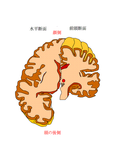
I took a look at the horizontal section and the frontal section.In a horizontal section, the top is the face and the bottom is the back of the head.?It's like looking through from the top of my head.
The basal ganglia are found in the medulla of the cerebrum as a large mass of gray matter.。
What is gray matter??It is a nerve cell.
In the first place, the surface layer of the cerebrum is occupied by gray matter over a thickness of 2 to 4 mm, and it is called the cerebral cortex.The reason it is called gray matter is that nerve cells are densely gathered and are grayish white.。
The area rich in nerve fibers is called the medulla.
The nerve fibers are covered with a myelin sheath, which is white because the myelin sheath is white. The medulla is also called white matter.
The basal ganglia, a cluster of nerve cells, was displaced from the gray matter to the white matter rich in nerve fibers.
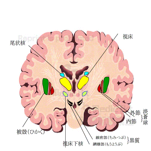
Then, what kind of basal ganglia (the name is from placing at the bottom of the cerebrum)??????
Caudate nucleus
Putamen
pallidum
These are the main part.besides these the substantia nigra of the midbrain and the subthalamic nucleus of the diencephalon are included in basal ganglia in some cases.
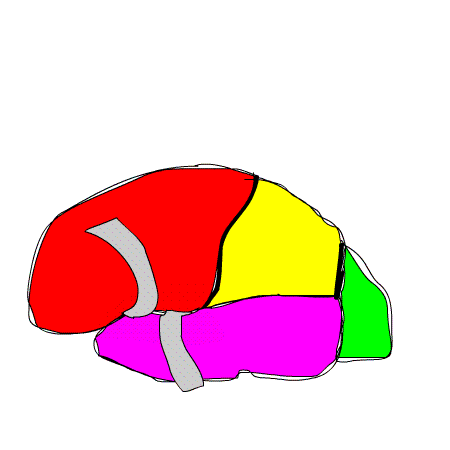
Let's expand the basal ganglia and take a closer look.In the animation below, red is the frontal lobe and purple is the temporal lobe.
Let's see the inside between lobes (lateral sulcus).First, you can see the area called insula.
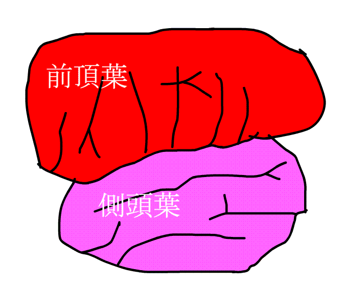
In animation,the left is the face and the right is backside of the head. This is the animation seeing from the side of the head.
At the back of the insula, the putamen can be seen, at the back of which there is a pallidum, and then the caudate nucleus appears.
There is an internal capsule in which several nerve fibers run between the caudate nucleus and putamen.
The thalamus is at the back of internal capsule. thalamus, internal capsule and caudate nucleus in the opposite appear.
The basal ganglia communicate with each other via nerve fibers.
It is a review.Basal ganglia is composed of the part below.
the striatum(caudateand putamen),globus pallidus (separeted to the outer segment and the internal segment) ,the substantia nigra(separeted to compact part and reticular part) ,subthalamic nucleus
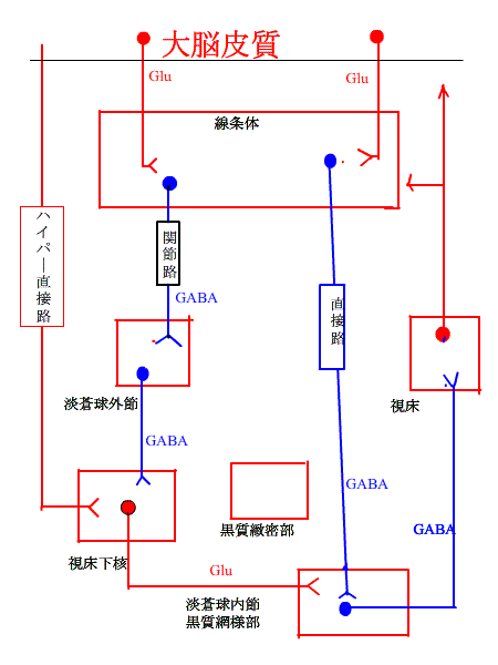
Stimulation is transmitted to the striatum from a wide area of the cerebral cortex via nerves.
As will be introduced later, stimuli are transmitted to the putamen via nerve fibers from the motor cortex of the cerebral cortex, somatosensory cortex, associative cortex related to cognitive function, orbitofrontal cortex related to emotion and cingulate cortex. Stimuli are transmitted from the thalamus and substantia nigra to the caudate and putamen .
It is shown in the animation of brain below.is represented by?the?blue line.?The stimulus starts from a round place, and is transmitted to Y.
On the contrary,in the red line,the stimulation is transmitted from the striatum which is the putamen and caudate nucleus?, respectively, to outer segment and the internal segment of globus pallidus and the subthalamic nucleus
In the animation on the right,stimuli are transmitted from the putamen, subthalamic nucleus, and caudate nucleus to the outer and inner segments of the pallidus.It is shown with a blue line.
Stimuli are transmitted from the pallidum to the subthalamic nucleus and thalamus.It is shown as the move from the red circle to the Y shape.
Stimulation is transmitted from the substantia nigra to the thalamus and from the subthalamic nucleus to the substantia nigra.
The relationship between the basal ganglia and the cortex is
| Cerebral cortex-> basal ganglia-> thalamus-> cerebral cortex loop circuit |
The basal ganglia has no direct connection to the spinal cord.?The nerves that move the body need to pass through the spinal cord, but the nerves that enter and exit the basal ganglia are unrelated to the spinal cord.?It is a basal ganglia deep inside the cerebrum that creates smooth and fine movements.
Now, I have to tell you a little difficult story.
The input and output shown in the animation above can be organized in two ways.
One is a direct path from the striatum to the globus pallidus internal segment or the substantia nigra reticular part.
The other is an indirect path that flows from the striatum to the globus pallidus external segment and the subthalamic nucleus and then to the globus pallidus internal segment or the substantia nigra reticular part.
The information of these nerves is a substance called GABA (r-amino acid) and is transmitted to the next nerve.?The GABA nerve is?shown by the?blue line in?the figure below?.?There are various nerves, but when the round part of this nerve becomes active,?a substance called GABA comes out from the shape of Y.?This is the nerve from the striatum to the outer segment of the pallidum, the inner segment of the pallidum and the substantia nigra, and from the outer segment of the pallidum to the subthalamic nucleus, from the inner segment of the pallidum and the substantia nigra to the thalamus.
| What is GABA??This has the effect of weakening nerve activity.?The nerve that receives this GABA becomes weaker when it conveys information next time. |
What is the red line? Expressed in Glu, when a substance called glutamine comes out of the Y-shape, it excites the next round sign of the nerve.
In the nerves that enter the cortex from the hyperdirect pathway and the thalamus, the excitatory substance is not Glu.
The activity of the thalamus is determined by whether the inner segment of the pallidum and the substantia nigra reticular part are weakened or stimulated and excited.
When the nerve activity of the thalamus becomes active, the motor area of ??the cerebral cortex is stimulated and the movement is accelerated.
On the contrary, when it is suppressed, the movement decreases.
Next, let's see the relationship between this hyperdirect pathway, joint path and direct path under normal conditions.
Please think while watching the animation below.
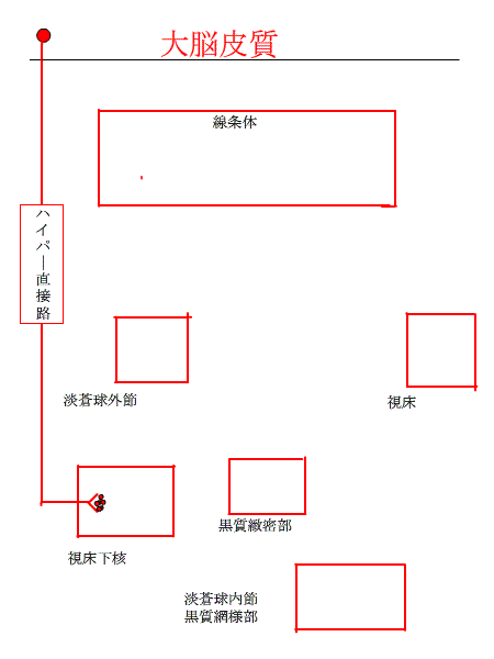
This is the conclusion. In mammals with basal ganglia, first, (1) signals from the cerebral cortex activate the hyperdirect pathway, and neurons in the pallidus inner segment and the substantia nigra reticular part. The neurons in the thalamus-cerebral cortex are suppressed and movement is reduced.
(2) Next, the signal through direct path becomes active, suppresses the activity of neurons in the pallidum inner segment and the substantia nigra reticular part.The neurons in the thalamus-cerebral cortex become active and the movement is accelerated.
(3) Finally, the signal through indirect pathway becomes active, the activity of neurons in the inner segment of the pallidum and the substantia nigra reticular part also becomes active.The neurons in the thalamus-cerebral cortex are suppressed, and movement is reduced.。
In other words, with this flow,exercise tends to be suppressed at all times,and unnecessary exercise is controlled.?In this term, we are trying to do the required exercise at the correct timing.
About Parkinson's disease
Before that, in the picture above, we didn't mention the compact part of the substantia nigra.?From this place, a substance called dopamine constantly comes out and acts on the places D1 and D2 of the Striatum.?The red line?represents the?nerve?that?activates it, and the?blue line?represents the?nerve?that?suppresses?it.?Dopamine has both function of activating and suppressing it.?In the anime, it is shown as a?black dot.?
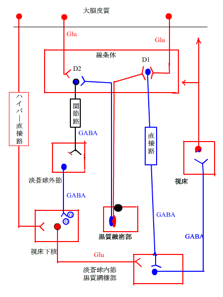
Look at the movement of Dopamine with black dots.
It works on D2 and D1, Ultimately, the nerves in thalamus excites in both direct and indirect ways.
Dopamine actively excites exercise in this neural circuit.
Parkinson's disease is caused that dopamine secreted from the substantia nigra reticular part is reduced.
In simple terms, when dopamine comes less, the body's movement is extremely decreased. Immobilization,inability of movement is caused by the lack of dopamine.
The tiny movements are caused by the lack of smoothness.
You can see the defference of tremor before and after the treatment called subthalamic nucleus stimulation method.
That's it for the basal ganglia.
?
There is one important thing
You could see the anterior limb and the posterior limb of internal capsule in the horizontal section of an anime。The inner capsule is a path of nerve fibers that connects the cerebral cortex and the basal ganglia from both sides.The inner?capsule?looks like a dogleg,and?from?the?corners to the posterior limb,nerve fibers run from the cerebral cortex to the spinal cord, and motor output fibers are mixed.
On the other hand, in the basal ganglia region, many blood vessels are distributed as in the animation below.。

These blood vessels are called perforators, which are branches of large blood vessels of the internal carotid , posterior communication, middle cerebrum, anterior cerebrum, and posterior cerebral arteries, which enter the tissues of the brain and are distributed in the interbrain and basal ganglia.
The Lenticulostriate arteries, which is a branch of the middle cerebral artery, controls the pallidum, putamen, and ?genu of internal capsule, often bleeds.This is why it is called stroke arteries.
| When bleeding in the middle cerebral artery affects the posterior leg of the internal capsule, the central part of the motor nerve is destroyed, resulting in hemiplegia and immobility of the limbs. |
The cerebral hemorrhage is likely to occur near the iternal capsule, and frequently occers at the putamen and thalamus.
Back to the first page. Jump to the Medicine for the child who learns by anime.