

Hearing
The basics of hearing are explained in "Medical Education for Kids for Children", with the title "What Can I Hear?"
The URL is http://www.skmc.jp/anime/child/ear/ear_index.htm.
Now let's think about how we catch the sound in our brain.
I brought the trailer to the front. I made it and felt difficult.I hope you find this interesting.
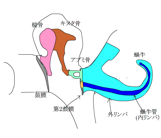
Vibration of the eardrum is transmitted to the perilymph of the inner ear through the ossicles.
Do you know the roots of the ossicle?
It is just a theory, but the ossicle was used to be jawbone when humans were still fish at ancient time.In the process of fish landing and evolving into humans,perhaps they needed to develop their hearing to survive. It seems that the bones of the chin were diverted into bones for listening to the sound.
Tsuchi → Hammer (the one that the rabbit holds on the moon) Kinuta → Tapping table for licking the leather. I thought Tsuchi and Kinuta as a pair. Abumi → Stirrup This is exactly the shape.
The role of the ossicles is to convey a slight sway of the eardrum into the cochlea. However,the inside of the cochlea is filled with liquid, and the "sway of the air → sway of the eardrum" is not transmitted with a little gentle force. To make matters worse, the cochlea is also a very delicate organ, so if a strong shaking (loud noise) is introduced, it will easily break.
It sounds hard to handle!
It is the ossicles that solves this difficult problem.
A masterpiece created by God, which strongly conveys even a slight sway of the eardrum to the cochlea, and protects the weak and delicate cochlea without further shaking!
It may be overstated, but this is a quite good precision mecanism.
The key words for these functions are "lever" and "area ratio" and a small muscle called the middle ear muscle (ossicle muscle).
There are joints between the "?Malleus" and "Incus", and between the "Incus" and "Stapes", each of which acts like a hinge on a door.
Here, the "Incus" is actually shorter than the "Malleus," so the structure here plays the role of a "lever."
In addition, the fact acts strong that the area of the eardrum is more than 10 times larger than the area of the outer window of the cochlea where the stapes are attached.
Imagine that.
There is a large membrane called the eardrum.
On the back of the eardrum, the “Malleus” and “Incus” are attached in a hinge-like structure, and the length of the “Incus” is shorter.
When the eardrum is pushed, this structure acts like a "lever," strengthening its power and being transmitted to the "Stapes".
The “Stapes” is connected to a small window that is less than 1/10 the area of ??the eardrum.?The pressure applied to the eardrum is amplified 20 to 30 times and transmitted to the inner ear.
Imagine a sharpened pencil here.
Put the tip of a sharp-edged pencil on the palm and push it a little from above,and even if the strength is small, it hurts a lot, isn't it ?
This is because the force applied to a large area (the bottom of a pencil) concentrates on a small area (the core of a sharp pencil), and a small force is transmitted as strong energy.
The same thing is happening in your ears.
The force applied to the eardrum is concentrated in the small "stapes" and becomes a strong energy.
And the ossicles are not just stuck together.
There are small muscles for sticking ossicles together.
This muscle automatically contracts (shrinks) when a loud sound comes in, and then suppress the movement of the ossicles.
This is a phenomenon called the "middle ear reflex", and because of this, it can protect a delicate cochlea when a loud noise comes in.
Next, let't talk about the inner ear.
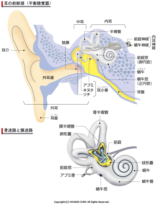
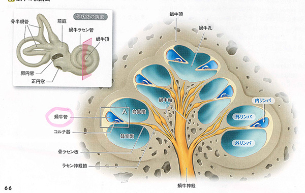
The inner ear consists of osseous labyrinth and a membranous labyrinth.
It plays the role of hearing and balance.
The nerve related to hearing is the cochlear nerve, and the nerve related to balance is the vestibular nerve.
These two nerves merge to form the vestibulocochlear nerve.
Quoted from wikipedia.
?
The bone maze refers to the cochlea, semicircular canals and vestibule.
?
The membranous labyrinth is the blue part in the picture above.
It is a soft,?membranous, closed tube?in the osseous labyrinth.?The endolymph is filled in it.?
The cochlear tube looks like osseous labyrinthis and is inside the cochlea.
The utricle and saccule are in the vestibule.
The semicircular canal is contained within the osseous semicircular canal.
They are much thinner than the osseous labyrinth and float in the perilymph away from the bone wall.?The picture below shows the membrane maze.?The gray one is the ossicle.?Endolymph fluid is contained in this membrane maze.
Quoted from gettingimages
Quoted from wikipedia
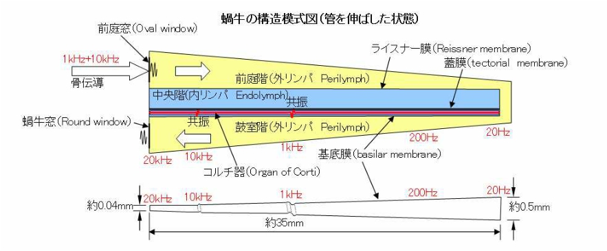
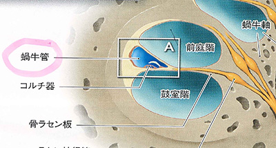
The figure above is a cross section of the cochlea seen from the top of the cochlea.?Quoted from wikipedia.?The cochlea is like a three-story tunnel.
A similar figure is shown below.
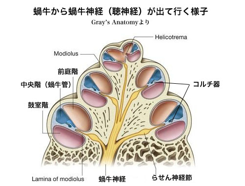
The cochlea is divided into the scala tympani, the?scala vestibuli and the scala media (cochlear duct).
Perilymph fluid flows in the scala tympani and the?scala vestibuli.?Endolymph flows in the scala media.
Next,?we will show?how sound waves are transmitted in?the?cochlea?.
Vibrations from the stapes are transmitted to the perilymph on the the?scala vestibuli.?The animation below shows the inside of the cochlea.?Oscillations of the perilymph go to the helicotrema, as indicated by the red arrow.
The sound waves decay from the top of the tympanic floor, descending and reaching the secondary tympanic membrane, as indicated by the blue arrow.
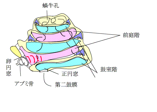
The triangular part shown in purple is the cochlear duct.
Both sides of the cochlear duct are closed.?A thin tube in the the?scala vestibuli connects with the sacculus.
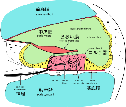
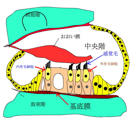
There is a receptor of sound inside the cochlea duct.It is the organ of corti shown below.?I enlarged the triangle part of the animation above.
When a sound wave is transmitted to the perilymph on the?scala vestibuli, the endolymph on the scala media also vibrates.
At this time, the membrane between the?scala vestibuli and the scala media is stiff and does not vibrate, causing the basement membrane under the organ of Corti to sway significantly.
Quoted by wikipedia
basement menbrane is located here
In the animation of the cross section of the cochlea shown below, the perilymph of the?scala vestibuli vibrates, causing the basement membrane of organ of corti in the scala media to vibrate.Therefore the sensory hairs at the tips of the inner and outer hair cells bend and stretche.?This mechanical stimulus opens and closes the mechanical demand channels of the sensory hair, generating action potentials.?This electrical signal is transmitted to the brain via nerves.
| The vibration of the basement membrane determines the pitch of the sound. |
There are two types of hair cells: inner hair and outer hair. Inner hair receives sound and outer hair regulates sound sensitivity. For humans, one cochlea has about 3500 inner hair cells and 12,000 outer hair cells. First, let's talk about inner hair cells .
At the base of cochlea near the fenestra vestibuli, where the sound waves first propagate, the basement membrane is narrow and stiff, so it tunes to a higher frequency (200000 Hertz). It becomes wider and softer toward the top of the cochlea,then it tune to lower frequencies (up to 100 hertz and below).
Perilymph oscillations best vibrate certain parts of the basement membrane that tune to that frequency. You can distinguish the pitch of the sound by the characteristics of the basilar membrane. See the figure below.
Next,Let's learn the way?the action potential is generated in?the?cochlear canal.
Originally, the electrolyte composition on the scala vestibuli and the scala media was very different.
The perilymph fluid flowing through the scala vestibuli and scala tympani has an electrolyte composition similar to extracellular fluid.
The endolymph fluid following to the scala media is similar to the intracellular fluid.
 This figure is the exanded cochlear canal.
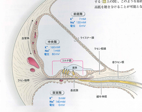
Na ion is 130 mM, K ion is 7 mM, and the potential is 0 mV on the scala vestibuli.?The Na ion is 1 mM, the K ion is 160 mM, and the electric potential is 80 mV in the scala media.?Clearly, K-ion is abundant in the scala media.
The sensory hairs of hair cells have mechanoreception channels which is opened by the basement membrane shaking.
In the endolymph, a high concentration of K ions flows into the open channels according to the concentration gradient.?This causes depolarization.
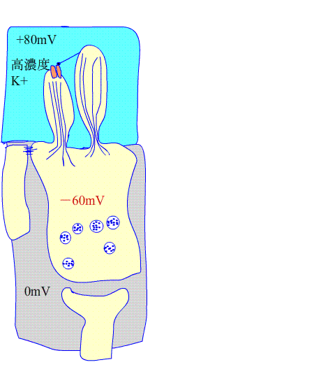
Please see the animation on the left.?The blue part is the scala media with endolymph.?Vibration of the basement membrane causes hair cells to bend.?This mechanical stimulus opens mechanoreceptive channels, allowing K ions to flow into the cell and causing a depolarization.?The -60mV rises up to around 0mV.?Then, the voltage-dependent Ca ion channel opens.?Many Ca in the perilymphions flow into the cells .
This changes the membrane potential, opening the round synaptic vesicles and secreting the transmitters within the vesicles.?This substance is glutamic acid.
The incorporated glutamate to the postsynapse excites afferent fibers, which is the nerves to the cerebrum, generate action potentials.?(The action potential will be explained later.)
About outer hair cells.
When outer hair cells are depolarized, that is, when these cells are excited, they increase the vibration of the basilar membrane, making it easier to tune the organ of Corti and the frequency of the sound, like inner hair cells.?To explain a little more, when the basement membrane vibrates by sound stimulation, the ear hairs on the heads of outer hair cells shift and move.it cause potassium ions to flow in and depolarize the cells. Furthermore, it vibrates the basement membrane largely.
In addition, the efferent fibers at the olivo‐cochlear bundle distributed in outer hair cells hyperpolarize outer hair cells in response to a command from the brain.?It also can suppress excitement.
In other words, when a sound is caught in the inner hair cells, when it is a small sound, the outer hair cells are excited and amplified to a loud sound.
When the sound is abnormally loud, the efferent fibers from the brain suppress the excitement and correct it into a soft sound.
| If the outer hair cells do not work, you can hear , but you will not be able to clearly understand the content of the story.。 |
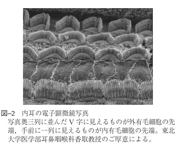
Dissection of outer and inner hair cells.
Quoted from Audiology Japan 59,161~169,2016
How is information,which is converted from sound waves into action potentials by the organ of Corti, processed in the brain ??The analog data called sound waves are converted to digital data by the Corti device.
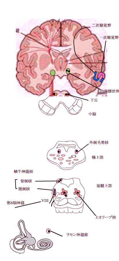
The simplest route is through four neurons into the auditory cortex of the cerebral cortex.?(1) First, primary neurons make synapses with inner hair cells and convert the information of hair cells into action potentials.?Axons extend from the spiral ganglion to the cochlear nucleus in the medulla oblongata.?② The axons of the cochlear nucleus intersect within the medulla oblongata, pass through the lateral lemniscus, and go to the inferior colliculus.?③ Axon extends further from the inferior colliculus to the medial geniculate body.?④ From here,it goes to the auditory cortex.
In the auditory nerve tract, all nerves to the cerebrum always pass through the inferior colliculus.
In addition, the auditory nerve path mostly intersect in the brain stem.?Even at the cerebral level, left and right communicate via the corpus callosum.
The auditory center has many collateral pathways.?For example, some way connect from the cochlear nucleus to the olivo‐cochlear bundle.?It also contacts the brainstem reticular activation system to excite the entire nervous system in response to loud noise.
The efferent fibers of the olivo‐cochlear bundle distributed in the outer hair cells run efferently from superior olivary nucleus?to the cochlear nerve ,which means they goes from the center to the ear.?It has a function of suppressing the activity of the cochlea, and protects the cochlea under strong sound.?Various routes are connected to the left, right and up and down.
Next, sound waves of various frequencies are converted into electrical signals (action potentials), transmitted through nerves, and go to the center.?At this time, the cochlear nerves are distributed corresponding to each frequency, and the nerves are aligned in the cochlear nucleus.?It's hard to understand, so please see the anime below. 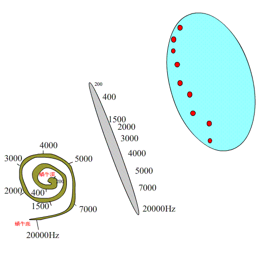
Next let's talk the auditory cortex.?It starts from the cochlear nucleus to the superior olivary nucleus?above the medulla oblongata, through the inferior colliculus of the midbrain, through the medial geniculate body, to the primary auditory cortex.
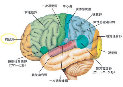 Roughly speaking, neurons that respond to low-frequency (approximately 100Hz) are on the dorsal side of the primary auditory cortex, and neurons that respond to high-frequency (approximately 20kHz) are inside of it. The neurons are arranged at the cerebral cortex in the gradation in order each frequencies.As shown in the figure on the left, the primary auditory cortex is the auditory cortex, which is located in the both sides of temporal lobes, and the auditory association area is around it, which regulates various sounds.?More nerves work in hearing than in vision.