

Points of this page With large wrinkles (sulcus), the cerebrum is divided into the frontal lobe, parietal lobe, occipital lobe and temporal lobe. The root is one. The surface of the cerebrum is called the cortex, and 90% of the cortex is made up of the neocortex. The neocortex is made up of 6 layers of organized cells.?Besides the neocortex, there are the paleocortex and the archicortex. In the neocortex, there is a place called the association area that collects various information.?It consists 30% of the total neocortex in human which is more than other creatures. |
Pushing the enlarged?neocortex?into the bones of the hard head?created?many?wrinkles ?on the surface of the brain?.
Next,?let's look at?wrinkles?.
In the anime above,?wrinkles?are called sulci.?The space between wrinkles and wrinkles is swollen, so it is called the gyrus.
These wrinkles increase the surface area of ??the cerebral hemisphere and increase the volume of the cortex.?The large wrinkle is the central groove.。
Please see the animation below.?The wrinkles of the central groove, lateral sulcus and sulcus parietooccipitalis divide the cerebrum into four parts.?The central groove separates the frontal lobe and parietal lobe.。
The sulcus parietooccipitalis separates the parietal and occipital lobes, and the lateral sulcus separates the temporal lobe.
Red is the frontal lobe, yellow is the parietal lobe, purple is the temporal lobe, and green is the occipital lobe.
Let's look at the brain from below.?I cannot see the parietal lobes.
From these animations, you see that the brain is divided into four parts with wrinkles (grooves) and bulges (gyrus) and large wrinkles.
Furthermore, when the brains are cut in the middle, the inside surface becomes visible.
Please see the animation below.
Here, a new blue part is visible. This is called the limbic system. This area is composed of the cingulum, the ?paraterminal gyrus, the cingulate gyrus, and the parahippocampal gyrus.(These words are difficult,take it easy.)
The gray area is the corpus callosum, which connects the left and right brains.
So what is the neocortex?
In the first place, the surface layer of the cerebrum is occupied by gray matter over a thickness of 2 to 4 mm, and it is called the cerebral cortex. The reason it is called gray matter is that nerve cells are densely gathered and are grayish white.
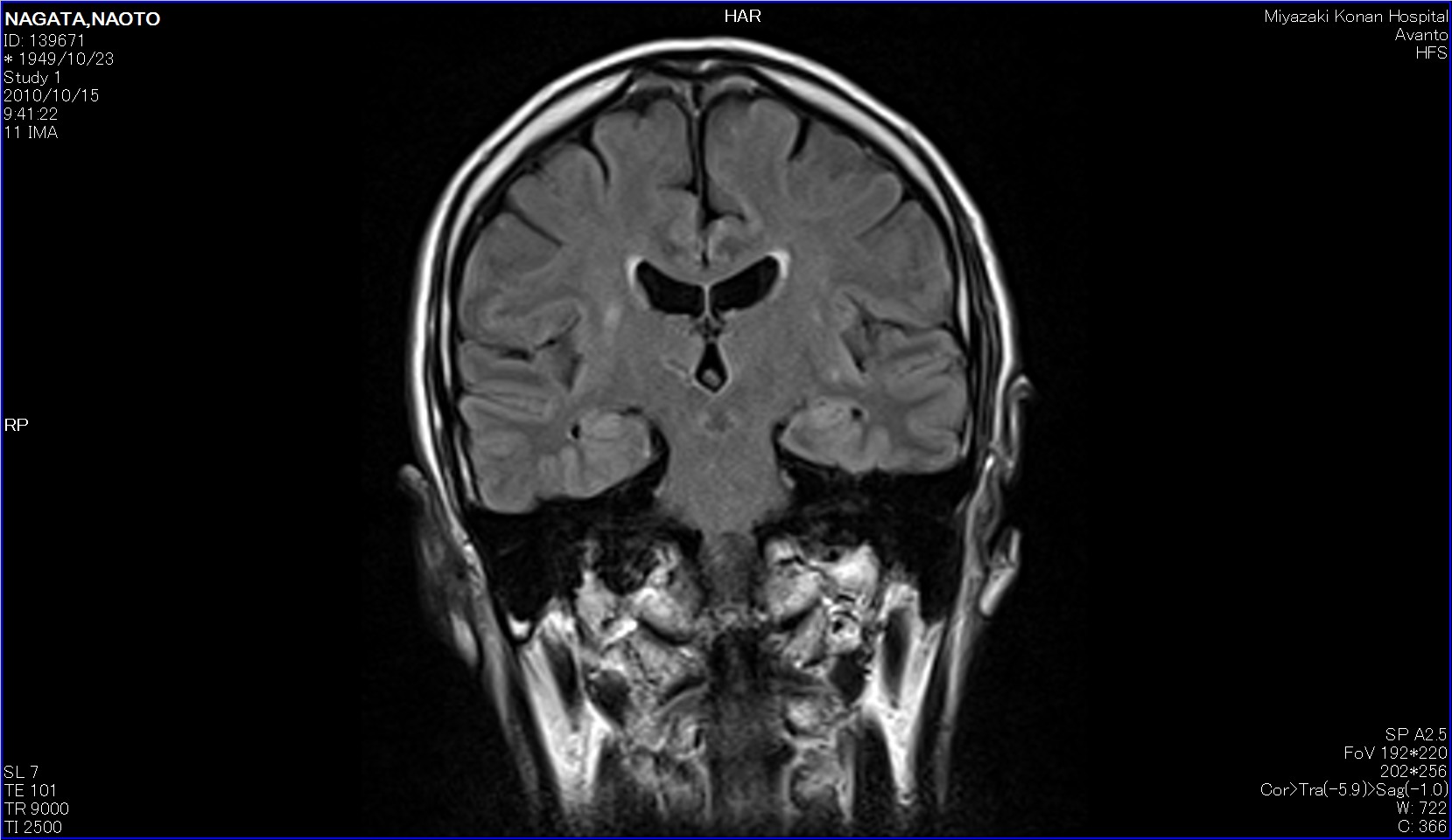
The picture on the left is a vertical section of the head taken by MRI.
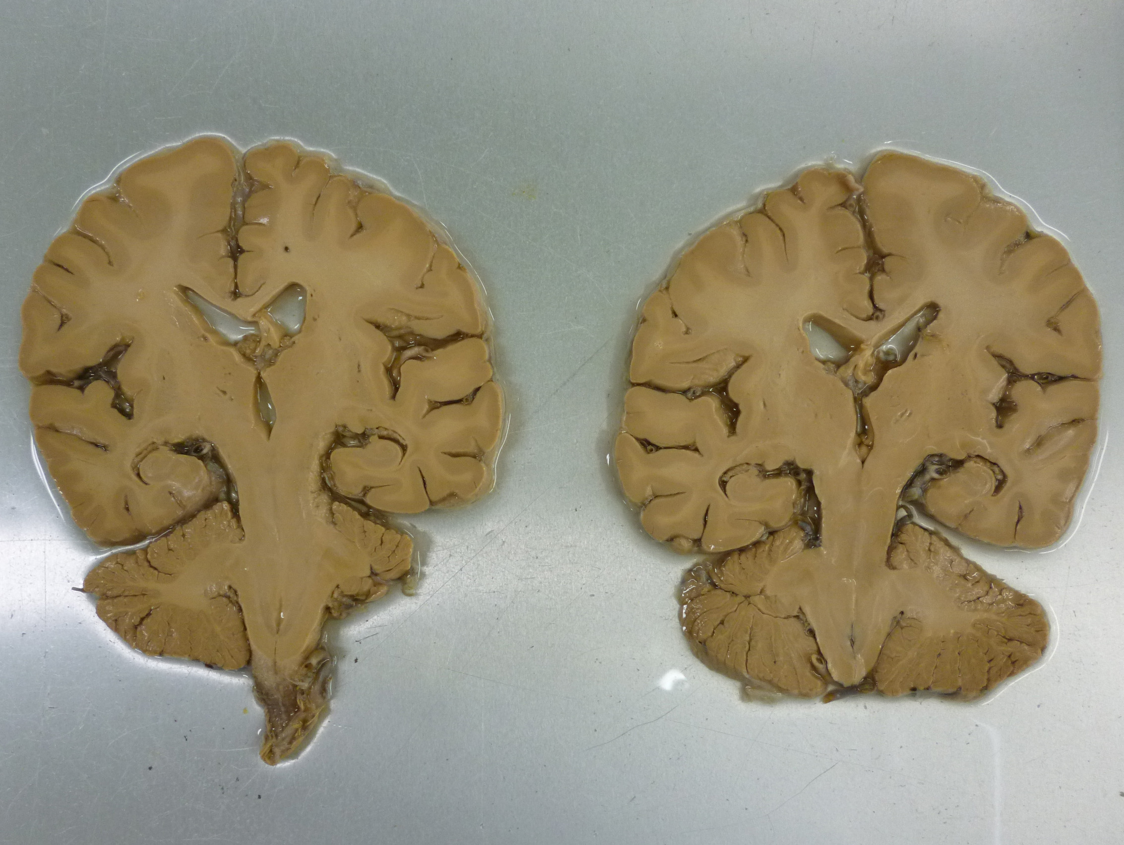
The picture on the right is an actual specimen of it.
Can the samples show the difference in color? Originally it is grayish white, but because it is a specimen, the outside is slightly brown and the inside is whitish. The cerebral cortex is the gray matter.
The white part inside is called the medulla. It is a place rich in nerve fibers.
The nerve fibers are covered with what is called a myelin sheath,and the nerve fibers are white because the myelin sheath is white.
By the way, in humans, 90% of the cortex is occupied by the neocortex.
There are about 14 billion neurons in the cortex, and they are divided into several layers by the method of staining the cells.
The neocortex is made up of 6 layers, each of which is lined up in order without mixing with other cells.?It is an evolutionarily new part of the cortical structure that occupies the surface and controls rational and analytical thinking and language functions.?So-called?lower animals ?tend to be?smaller and?higher animals?tend to be larger.
Besides this, there are the paleocortex and the archicortex. These are made up of 3-5 mixed layers。
The picture below shows a 6-layer structure.
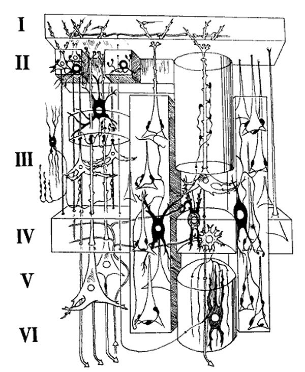
quoted from http://blog.livedoor.jp/subbody/archives/7716115.html
The name of the layer structure is number one layer being a molecular layer, and then an outer granular layer, an outer pyramidal cell layer, an inner granule layer, an inner pyramidal cell layer and a multiform layer of isocortex.
Similarly, it has a 6-layer structure.

http://detail.chiebukuro.yahoo.co.jp/qa/question_detail/から引用
What is the?Brodmann Area?
Focusing on a tissue staining method developed by Franz Nissl that stains different types of cells in the brain, Corbinian Broadman used it to analyze?human brain in 52 different areas in?1909.?He created this division that is now called Brodmann Area.
The division created by Corbinian Broadman was very accurate for many brain areas, such as the division of areas 17 and 18 in the visual cortex.
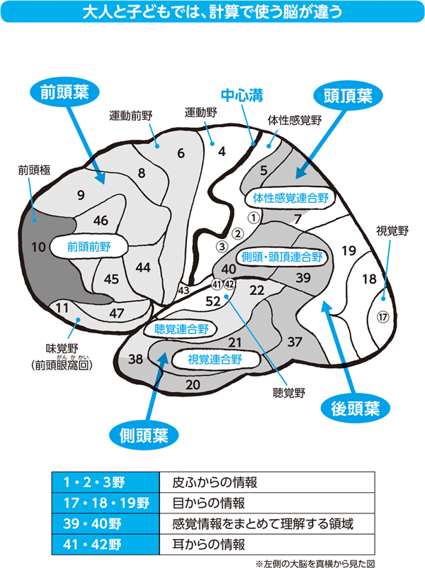
Quoted from http://diamond.jp/articles/-/94610