

| There are less animation in this page.I am sorry if you are less interested.This part of brain is not completely understood yet. |
By the way, people have lived since time immemorial, receiving many stimuli from the environment.
From the era of Aristotle, the five senses have been sight, hearing, taste, touch and smell, and getting the stimuli around them. Currently, the skin sensation typified by tactile sensation is distinguished from pain sensation, touch / pressure sensation , temperature sensation, cold sensation, and vibration sensation, and in addition, position, kinesthetic sensation, and balance sensation are also regarded as type of sensation.
In the cortex of the cerebrum, which we talked about in No. 7, there are centers corresponding to each cortex, roughly classified into motor cortex, sensory cortex, auditory cortex, and visual cortex.
Here, I will explain the association field.
Unlike other animals, humans have developed a neocortex area called the association area, which excludes the motor, sensory, auditory, and visual areas described above.
In humans, the myelination of nerve fibers in the neocortical region (from unmyelinated to myelinated nerves) is made the last in the brain.
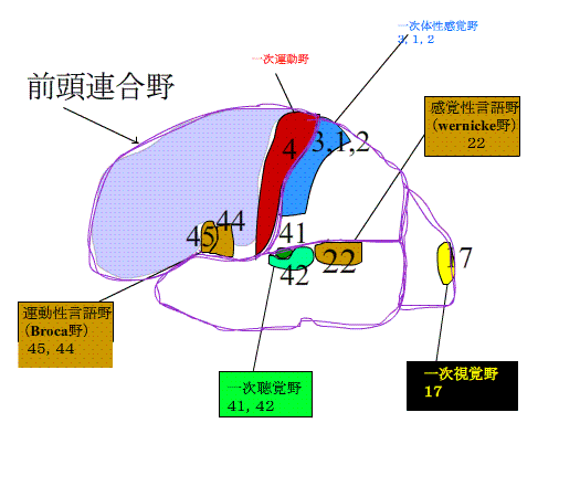
There are three allied fields. The?parietal association cortex, the parietal association cortex, and the frontal association cortex.
| The role of the association area is to receive and integrate the sensory information from the visual, auditory and somatosensory areas. Also, it makes an action plan based on sensory information and memory. |
In the "Structure of the brain 1" on this website, it was stated that the brain cells in the neocortex are 6 layers. In the middle of the occurrence, there is always one time when all 6 layers exist. After that, in the motor cortex, the 4th layer (granular cell layer) becomes weak, almost disappears, and 5 layers of pyramidal cells develop.
The sensory area has four layers developed, and sensory information is entered in this layer.
On the other hand, in the neocortex other than the sensory and motor areas, the first to sixth layers are almost evenly distributed.
The formation of insulating material around nerve fibers occurs more slowly in this neocortex than in the sensory and motor cortex.
It also exists between the sensory and motor cortex and has the characteristic of becoming wider as the animal becomes higher organism. This is the Association Field.
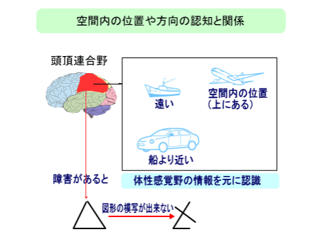
Disorders of parietal association cortex
A patient with a disorder of the right parietal lobe drew the right picture imitating the left picture.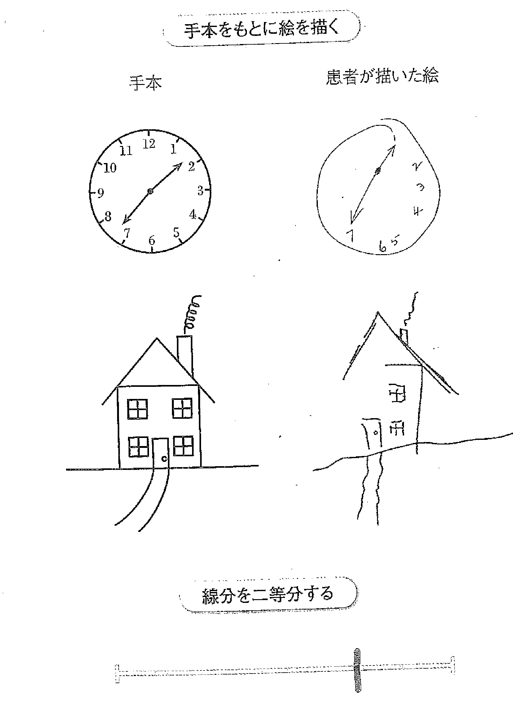
Neither number of the clock or the building on the left side are drawn.
When a patient bisects the horizontal line, it is biased to the right.
This is the symptom of the hemispatial neglect.
In addition, as shown in the picture below, patient cannot tell the location of a ship, car, or airplane . It seems to be due to an obstacle in the entire parietal lobe.
Temporal association cortex disorders
object agnosia Despite patient can see, they can not tell what they are. |
prosopagnosia Despite patient looks at person and celebrities they know, patient can't tell who they are. |
acoustic agnosia Patient can hear,but can not tell what they are. |
amusia Patient can not understand the melody or rhythm of music.。 |
Disorders of the frontal association cortex
The frontal cortex is concerned with the ability to set goals and plans and accomplish them effectively.
It is also related to personality and sociality. It controls emotion.
Patient with obstacle of the frontal association cortex can't flex your mindset, and can't get things done, one after another. The emotions are out of control,and personality changes to be whimsical, insidious, and arrogant . |
About motion
Humans achieve various purposes by making actions and working around them.
We can feel such as heat, cold, and pain, can see with eyes and listen with ears. Sensation is that specific organs catches sense and recognize external stimuli (sensory receptor). The senses are roughly classified as follows.
(1) Somatosensory: Somatic sensation is a combination of superficial sensation(cutaneous sensation) and bathyesthesia. There are tactile sensation, pressure sensation, sensation of warm, sensation of cold, and sensation of pain. Bathyesthesia include kinesthetics and deep pain.
(2) Visceral sensation: organic sensation (such as nausea) and visceral pain.
(3) Special sensations: visual sense(sight),auditory sense (hearing), sense of taste, sense of smell , vestibular sense (balance).
1. First of all, human beings consider the external world and the internal state of the body, and select the action to achieve the purpose according to the situation.
2. Assemble the movement patterns required to achieve that action. What kind of exercise, in what order and at what timing? (programming)
3. body actually does exercises based on that program.
4. Check and correct the exercise performed and the results of the exercise.
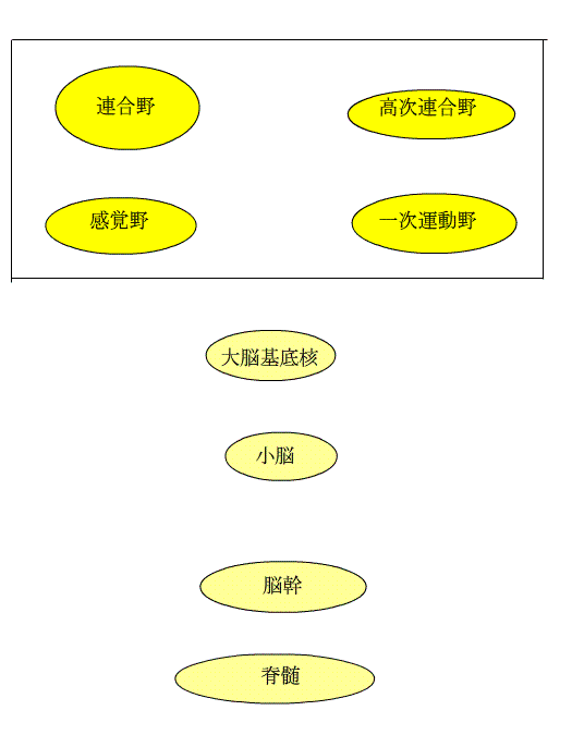
Programming, execution, and regulation of movements are performed by the cerebral cortex motor area, basal ganglia, cerebellum, brain stem, and spinal cord , as shown in the figure below . These are the five motor centers.
Control of movement
In the cerebrum, there are multiple motor cortex (premotor cortex, supplementary motor area, presupplementary motor area, and cingulate motor area), which are higher-order motor related areas.It receives various sensory and cognitive information from the sensory and frontal association areas. Obstacles in this area do not cause paralysis but impair movement.
For the primary motor cortex, see Chapter "Role of the cerebral cortex." When this area is damaged, the movement of the muscles corresponding to a certain cortex is lost and paralysis occurs, as in the horncrew dwarf .
Three characteristics of the motor area
1. Signals for movement flow from the cerebral cortex motor area, which is a combination of the higher-order association area and the primary motor area, surrounded by a square, to the brain stem and spinal cord below. In other words, like a building, the department is clearly divided into the motor cortex, brain stem, and spinal cord, and no signal flows upward. It is called a layered structure. In the animation on the left, the path from the primary motor cortex to the brain stem is called the?tractus corticobulbaris. The path from the primary motor cortex to the spinal cord is the tractus corticospinalis. Both go through the brainstem.This is explained in the Chaptar of the spinal cord and the pyramidal tract.
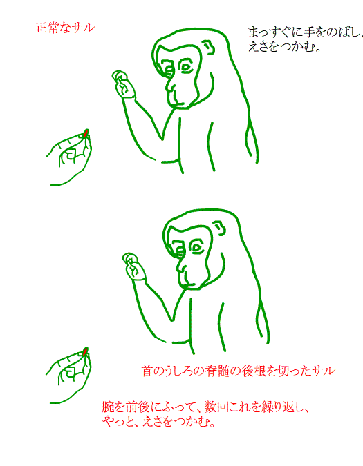
2.There are areas such as the face, upper limbs, and lower limbs in each motor center. For example, between motor centers, areas related to the upper limbs in motor area connect to the upper limbs motor neurons of the spinal cord but it never connect to the lower limbs neurons. This is called somatotopic organization.
3. Each motor center contains sensory information. The spinal cord has information felt by muscles, tendons, and skin. The brainstem and cerebellum also have balance sensory informationin addition to somatosensory information. In the motor area, information such as sight and hearing is also input from the sensory association area.
The spinal cord and brain stem have motor neurons that send output to skeletal muscle. This path is called the final common path, because commands are always sent from motor neurons to muscles in all movements. Also, the spinal cord and brain stem have circuits called reflexes . The brainstem regulates the spinal cord through neural circuits to the spinal cord, and controls posture and gait.
Regarding No. 2, for example, a monkey with damage of the bottom of spinal cord cannot move smoothly.This means the somatotopic organization is broken.
Please see the anime below.
The animation below shows the premotor and supplementary motor areas of the?higher-order motor related areas. Signals from the frontal association area and various sensory association area enter the premotor and supplementary motor cortex and are sent to the primary motor cortex and spinal cord.
Electrical stimulation of the higher-order motor related areas causes movement. In the supplementary motor area, there is area connected to the movement of the face, upper limbs, and lower limbs from front to back. In the premotor cortex, there are area for movements of the lower limbs, upper limbs, and face from the inside to the outside.Compared to the primary motor cortex, strong stimuli are required to induce movement, and movement is also complicated. When the higher motor area is impaired, the person is not paralyzed but he cannot exercise well.
When the supplementary motor area is impaired,the person speaks less and does not move initiatively.he is able to move, but is not willing to do. He can not hit the instrument with a certain rhythm or move your fingers and shoulders or elbows at the same time.
Characteristics of higher motor areas different from primary motor areas
The higher motor areas works more with complex ordered exercises than with simple exercises. The supplementary motor cortex is more active when it exercises based on information stored in the brain the premotor cortex. The premotor cortex is more active when it exercises based on visual information.
The higher motor areas works when you are preparing for the next exercise or when you think of a complex sequence of exercises in your head. It is recently figured out the pre-motor area is active when you see the movement of the other person.The pre-motor area seems to be related to understanding movements and communicating with other people.
